Monday- Friday 8:30 am - 5 pm
  Contact : (954) 454-1066

Identifying Best Candidates for Bexarotene Therapy in Cutaneous T-Cell Lymphoma: Role of APOA5, APOC3 Genotypes

Share this content:
The majority of the patients had mycosis fungoides, the most prevalent form of cutaneous T-cell lymphoma.

According to results of a retrospective, observational, noninterventionalist, multicenter Spanish case-series study designed to evaluate the role played by genetic polymorphisms in bexarotene-treated patients with cutaneous T-cell lymphoma (CTCL), screening of the APOA5 and APOC3 genotypes in these individuals may be a useful tactic for estimating triglyceride concentrations. Findings from the current analysis were published in JAMA Dermatology.

The investigators sought to examine the link between genetic polymorphisms of apolipoprotein genes APOA5, APOC3, and APOE, and to evaluate the severity of hypertriglyceridemia among patients with CTCL receiving bexarotene therapy, as well as to optimize patient selection for treatment with bexarotene, based on the expected adverse effect profile.

Continue Reading Below

The case series was performed at 12 university hospital dermatology departments in Spain between September 17, 2014, and February 17, 2015. A total of 125 patients with a confirmed diagnosis of CTCL who had received bexarotene for ≥3 months were enrolled in the study. Ultimately, 9 individuals were excluded because of missing analytic triglyceride data; thus, the final study group comprised 116 patients.

Data on demographic and cardiovascular risk factors were obtained for all participants. In addition, a complete blood analysis was performed, including a lipid profile, along with a genetic analysis from a saliva sample. The primary study outcome was the maximal triglyceride levels that were reported in association with the minor alleles of the polymorphisms being analyzed.

The mean patient age was 61.2±14.7 years; 59.5% of the participants were men, and 73.2% had mycosis fungoides, the most prevalent type of CTCL. While undergoing bexarotene treatment, 82.8% (96 of 116) of the participants developed hypertriglyceridemia, which was considered to be severe or extreme in 8.3% (8 of 96) of these patients.

Those individuals who carried minor alleles of the polymorphisms did not demonstrate any significant differences in baseline triglyceride levels. After bexarotene treatment, however, carriers of ≥1 of the 2 minor alleles of APOA5 c.1131T>C and/or APOC3 c.*40C>G had significantly lower concentrations of triglycerides compared with noncarriers of these minor alleles (241.59±169.91 mg/dL vs 330.97±169.03 mg/dL, respectively; P =.02), and were thus less likely to experience severe hypertriglyceridemia.

The researchers concluded that the findings from this study suggest that APO polymorphism investigation might prove beneficial in the clinical evaluation of bexarotene-induced hypertriglyceridemia in patients with CTCL and identification of the most suitable candidates for this treatment. Future studies aimed at exploring the sensitivity of these polymorphisms for predicting adverse outcomes to recognize which patients might need to discontinue treatment, or which will experience pancreatitis are warranted.

Follow @DermAdvisor

Reference

Cabello I, Alia P, Pintó X, e al. Association of APOA5 and APOC3 genetic polymorphisms with severity of hypertriglyceridemia in patients with cutaneous T-cell lymphoma treated with bexarotene. JAMA Dermatol. 2018;154(12):1424-1431.

Read More

Actinic Keratoses: Field Cancerization and Photodynamic Therapy

This article originally appeared here.

 Each month, The Clinical Advisor makes one new clinical feature available ahead of print. Don’t forget to take the poll. The results will be published in the next month’s issue.

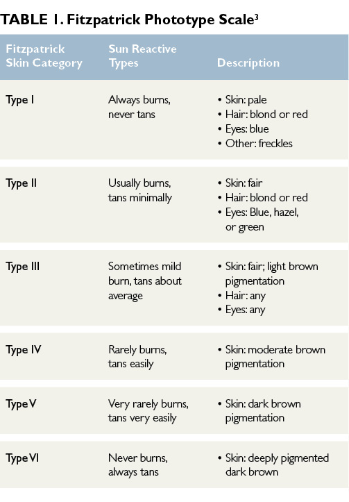
Actinic keratoses (AKs) are one of the most frequently encountered skin lesions in clinical practice.1,2 AKs are precancerous, focal, sun-induced areas of abnormal proliferation of atypical keratinocytes confined to the lower layer of the epidermis.1,2 Often referred to as solar keratoses, these lesions commonly occur in areas with chronic and cumulative sun exposure such as the face, ears, neck, dorsal forearms, hands, and scalp.1,2 The highest incidence of AK is seen in middle-aged to elderly men with chronic and sustained ultraviolet (UV) radiation exposure over their lifetimes. AKs are more prevalent in fair-skinned individuals and those categorized as Fitzpatrick skin types I and II (Table 1)3; the Fitzpatrick phototype scale is a numerical classification used to estimate the response of different types of skin to UV light.3

Click to enlarge

Etiology/ Histology

The main mechanism of AK formation is prolonged sun exposure and cumulative exposure to UV radiation.4 AKs result from the adverse effects of UV-A and UV-B light radiation on keratinocyte DNA.2,5 UV radiation induces cellular mutations and results in the formation of atypical keratinocytes.2,5 Mutations of genes may cause impaired apoptosis, uncontrolled cell proliferation, and progression to invasive squamous cell carcinoma (SCC).1,6 Although most AKs remain superficial and confined to the epidermis, some lesions will penetrate the reticular or papillary dermis and differentiate into SCC, especially when multiple AKs are present.1,6

Which of the following is the least effective topical therapy for actinic keratoses?

Clinical Presentation

The classic presentation of AKs is a gritty macule or small papule, varying in size, with an erythematous base and an overlying scaly white patch that is rough to palpation.2 Although AKs are commonly asymptomatic, patients may report occasional burning or lesion tenderness. Immunocompromised patients, specifically organ transplant recipients receiving treatment with immunosuppressive medications, are at increased risk of developing AKs.1,6 AKs may spontaneously regress, completely resolve, persist, or progress to SCC, particularly in the presence of multiple AKs.2 Clinically it is difficult to predict the course of AKs, and no method exists for predicting lesions that will evolve into invasive SCC.5 The risk of AKs transforming to SCC is minimal (1%)1; however, current guidelines suggest treating all AKs.4

Diagnosis

Diagnosis generally is made by visual inspection and palpation. Keratoacanthoma, SCC, basal cell carcinoma, and seborrheic keratosis lesions may have a similar presentation to AKs and must be considered in the differential diagnosis.2 Characteristic history and examination findings are usually adequate for diagnosing AK, but dermoscopy, if available, is recommended if the clinical exam findings are not typical of AK. A strawberry pattern, consistent with undulated vessels surrounding hair follicles filled with yellow keratotic plugs on an erythematous background, is characteristic of AK lesions on dermoscopy. Skin biopsy is performed if the clinical exam and dermoscopy findings are not typical of AK. Any clinically suspicious lesion — including lesions with increasing tenderness, rapid enlargement, or those recalcitrant to initial therapies — warrants skin biopsy for a definitive diagnosis.2     

Continue Reading Below

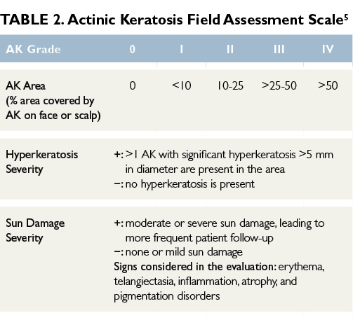
Surrounding the visible and palpable AKs lesions are skin areas prone to sun damage that are more likely to develop into clinical AKs or other sun-related cancers.5 Subclinical, nonvisible sun damage is known as field cancerization.5 Determining the severity of AKs and the extent of surrounding field damage can be challenging. Many AK grading tools use visible isolated lesion counts to assess the severity of the AKs, but they lack grading of the surrounding sun-damaged skin. The Actinic Keratosis Field Assessment Scale (AK-FAS) has been developed and tested as a relevant and reproducible assessment of AKs in clinical practice and aids in the grading of the severity of AKs and the field area damaged by UV radiation.5 Utilizing the scale can standardize a diagnosis, aid in selecting the most appropriate treatment, and provide an objective method to assess treatment response (Table 2).5

Click to enlarge

Read More

Unique Presentation of SJS in a Female Patient Treated With TMP-SMX

This article originally appeared here.
Share this content:
Stevens Johnson syndrome (SJS) and toxic epidermal necrolysis (TEN) often presents 8–12 days after drug exposure

A recently published case report describing a 27-year-old female patient with a history of polycystic ovary syndrome discusses a rare presentation of Stevens Johnson syndrome (SJS) involving vaginal pain and mucocutaneous desquamation following re-exposure to trimethoprim-sulfamethoxazole (TMP-SMX).

The patient, who presented to the clinical office complaining of dysuria, was diagnosed with a urinary tract infection and was prescribed TMP-SMX. After taking 4 doses of her medication, she presented to the emergency department (ED) complaining of left labial swelling and pain. She was diagnosed with left vulvovaginitis and was told to discontinue TMP-SMX since her urine cultures were negative. Two days later, the patient returned to the ED complaining of worsening vaginal pain and vulvar lesions but was discharged with a sitz bath and supportive care and urged to follow-up at the clinical office. At her follow-up appointment, she was reinitiated on TMP-SMX due to  “concerns of vulvar cellulitis with possible underlying abscess.”

Continue Reading Below

“After she received 2 doses of TMP-SMX, the patient had new lip swelling, periorbital swelling, facial swelling, and formation of thick white plaque on her tongue and mouth,” the study authors reported. They added, “She also developed an erythematous papular pruritic rash on her hands, arms, soles of the feet bilaterally and papules were noted on the legs (up to thighs), feet, and abdomen.”

The study authors also noted that the patient experienced early vesicle formation, skin sloughing, significant erythema and edema in her genital region, a fever of 102°F, plaques on her tongue, and a painful maculopapular skin rash. The patient was diagnosed with SJS and transferred to the burn unit/ICU where she received intravenous immunoglobulin, supportive care, as well as wound care. The patient was discharged 11 days after being admitted to the hospital.

In their review, the study authors discussed several interesting features of this patient case, including the quick onset and development of SJS (6-12 hours vs 6 days to 2 weeks) as well as the unique presentation of the patient (vaginal pain/ mucocutaneous desquamation). They concluded, “This report shows that although SJS is a rare diagnosis, providers should consider SJS as a possible differential diagnosis as a cause of vaginal lesion after exposure to drugs.”

Follow @DermAdvisor

Reference

Mergler R, Chuang M; Stevens Johnson Syndrome with Vaginal Pain and Lesions as Initial Presentation. Am J Case Rep 2018; 19:1519-1521 DOI: 10.12659/AJCR.912123

Read More

Switching Therapeutic Class in Psoriasis Linked to Better Treatment Efficacy

Share this content:
The investigators sought to describe switching patterns in dermatologic real-life daily practice among patients with psoriasis.

Among patients with psoriasis who require a switch in treatment, a change of therapeutic class appears to be more effective than a switch to an agent in the same therapeutic class, according to the results of a noninterventional, retrospective, multicenter analysis conducted at 11 volunteer dermatology centers in France. Findings from the study were published in Dermatologic Therapy.

The investigators sought to describe switching patterns in dermatologic real-life daily practice among patients with psoriasis. Efficacy was defined as achievement of ≥75% improvement in Psoriasis Area and Severity Index (PASI 75) score, which was evaluated between 12 and 16 weeks after initiation of a new therapy. Patients enrolled in the study included those who were seen in routine daily practice and were being treated with infliximab, etanercept, adalimumab, or ustekinumab. A standardized case report form was filled out for each participant, which included the patient’s characteristics and clinical details, including all prior treatments.

Continue Reading Below

A total of 1157 patients were included in the study, of whom 29.9% (346 of 1157) required a switch in treatment. The mean duration of treatment before the switch was 529.2 days for ustekinumab (range, 121-978 days), 450 days for etanercept (range, 13-2100 days), 431 days for adalimumab (range, 26-1729 days), and 424.2 days for infliximab (range, 4-1977 days; P =.21). When the first-line biologic agent was administered concomitantly with methotrexate, the mean duration of treatment was 530 days (range, 4-2100 days) compared with 439 days (range, 54-1638 days) when the biologic was administered as monotherapy (P =.14).

Switch in biologic agent, which was attributed to lack of efficacy, loss of therapeutic response, or adverse events, was considered effective in 71.9% of patients and ineffective in 28.1% of patients. Notably, switch efficacy was not known for 18 patients because of relocation or missing data.

The results provide a clearer understanding of the profile of patients with psoriasis who require a switch in their therapy, as patients were mostly men, were approximately 40 years of age on initiation of their first biologic agent, and were overweight (mean body mass index, 28 kg/m2). In addition, almost half of all individuals who switched had confirmed psoriatic arthritis.

The investigators concluded that the findings from this study are reflective of the high rate of comorbidities among patients with psoriasis and emphasize the role played by dermatologists in screening comorbidities among patients with psoriasis who require systemic therapy. This role is even more critical in the subpopulation of patients who require a switch from their first-line biologic therapy, regardless of the reason.

Follow @EndoAdvisor

Reference

Dabouz F, Khemis A, Barbe C, et al; Resopso network. Factors associated with successful switching between biologic therapies for the treatment of psoriasis in daily dermatological real-life practice: the Resoswitch study [published online November 28, 2018]. Dermatol Ther. doi: 10.1111/dth.12789

Read More

Increased Risk for Incident Thyroid Disease in Psoriasis

Share this content:
An endocrinology consultation should be considered in patients with psoriatic disease who present with thyroid symptoms.

Patients with psoriatic disease have an increased risk for thyroid disease, including hyperthyroidism, hypothyroidism, thyroiditis, and certain autoimmune thyroid conditions such as Grave’s disease and Hashimoto’s thyroiditis, according to study results published in the Journal of the American Academy of Dermatology.

The investigators identified a total of 13,266 patients with psoriatic arthritis (the psoriatic arthritis group), 149,576 patients with psoriasis alone (the psoriasis group), and 162,842 controls who did not have psoriasis (the control group) from data collected between January 1, 2000 and December 31, 2012. Mean patient age in the psoriasis, psoriatic arthritis, and control arms was 45.11±20.09, 43.17±17.72, and 44.95±19.91, respectively (P <.0001); women comprised 46.21%, 37.83%, and 40.65% of the groups, respectively (P <.0001). A significantly higher percentage of the patients in the psoriasis and psoriatic arthritis arms had hypertension, type 2 diabetes, and hyperlipidemia compared with patients in the control group (P <.0001).

Continue Reading Below

Compared with the control group, the psoriatic arthritis and psoriasis groups had an elevated risk for incident hyperthyroidism (adjusted hazard ratio [aHR] 1.32; 95% CI, 1.07-1.65; P <.05 and aHR 1.22; 95% CI, 1.11-1.33; P <.0001, respectively) and Grave’s disease (aHR 1.38; 95% CI, 1.07-1.79; P <.05 and aHR 1.26; 95% CI, 1.131.41; P <.0001, respectively).

Moreover, both the psoriatic arthritis and psoriasis groups also had an increased risk for incident hypothyroidism (aHR 1.74; 95% CI, 1.342.27; P <.0001 and aHR 1.38; 95% CI, 1.231.56; P <.0001 respectively) and Hashimoto’s thyroiditis (aHR 2.09; 95% CI, 1.343.24; P <.01 and aHR 1.47; 95% CI, 1.181.82; P <.01, respectively) compared with the control arm.

A major study limitation was the lack of available data on psoriasis severity. In addition, in order to avoid overestimating the risk for incident thyroid diseases, patients with psoriasis diagnosed prior to 2000 were excluded from this study.

The investigators concluded that the findings demonstrate that an endocrinology consultation should be considered in patients with psoriatic disease who present with thyroid symptoms, particularly patients with psoriatic arthritis.

Follow @DermAdvisor

Reference

Wang S-H, Wang J, Lin Y-S, Tung T-H, Chi C-C. Increased risk of incident thyroid diseases in people with psoriatic disease: a cohort study [published online December 5, 2018]. J Am Acad Dermatol. doi:10.1016/j.jaad.2018.11.049

Read More

Skin Changes, Risk for Severe Acneiform Eruptions Seen With EGFR Inhibitor Therapy

Share this content:
Oily skin was a risk factor for severe acneiform eruption.

Instrumental evaluation has demonstrated rapid inflammatory changes of the skin associated with epidermal growth factor receptor (EGFR) inhibitors and revealed that the presence of oily skin is a risk factor for severe acneiform eruption (AfE), according to the results of a multicenter, observational study conducted at the National Cancer Center Hospital, Tohoku University Hospital, and Mie University Hospital, all located in Japan. Findings from the analysis were published in The Journal of Dermatology.

Recognizing that EGFR tyrosine kinase inhibitors (TKIs) and anti-EGFR antibodies are often linked with the development of various dermatologic adverse events, and that the precise skin changes and risk factors for severe AfE remain unclear, the investigators sought to elucidate useful parameters for early sensitive detection of skin changes by EGFR inhibitors.

Continue Reading Below

Skin surface hydration, transepidermal water loss, skin surface lipid levels, and erythema/melanin index were all measured serially for 2 weeks among 19 people treated with the EGFR-TKI afatinib/erlotinib and for 8 weeks among 20 people treated with the anti-EGFR antibody cetuximab.

Transepidermal water loss levels of the cheek were already elevated 7 days following initiation of afatinib/erlotinib therapy in patients who had AfE grade 2 or more compared with prior to therapy and compared with patients with AfE of grade 1 or less. Further, in cetuximab-treated patients with AfE, skin surface hydration on the cheek in those with grade 2 or more AfE decreased significantly compared with hydration in those with grade 1 or less AfE at weeks 2 and 6 (P <.05). Baseline skin surface lipid levels and erythema index on the cheek of patients with grade 2 or more AfE were significantly higher than in those with grade 1 or less AfE (P <.05).

The investigators concluded that the major limitation of this study was the small sample size, particularly when performing logistic regression analysis.

The results of the current study revealed that AfEs resulting from EGFRIs develop subclinically as early as the first week of treatment in patients receiving EGFR-TKIs. In contrast, in patients who are treated with an anti-EGFR antibody, AfEs caused by EGFR inhibitors are observed during the second week of therapy. High skin surface lipid levels prior to EGFR inhibitor therapy and oily skin are both risk factors for severe AfE.

Follow @DermAdvisor

Reference

Kikuchi K, Nozawa K, Yamazaki N, et al. Instrumental evaluation sensitively detects subclinical skin changes by the epidermal growth factor receptor inhibitors and risk factors for severe acneiform eruption. J Dermatol. 2019;46(1):18-25.

Read More

Protecting Your Practice From Data Breaches: An Expert Interview

This article originally appeared here.
Share this content:
Passwords should not be visible to the public

The privacy of patient data is protected by the Health Insurance Portability and Accountability Act (HIPAA) and the 2009 Health Information Technology for Economic and Clinical Health Act.1 However, the years between 2010 and 2013 saw data breaches involving at least 29.1 million patient records, and the continuing transition in healthcare to electronic health records (EHRs) is likely to increase these breaches.1

A recent study1 analyzed all breaches in health information reported to the US Health and Human Services Office for Human Rights from 2010 to 2017 and found that breaches increased almost every year during the study period. Analysis of 2149 breaches, comprising a total of 176.4 million records revealed that, although the largest number of breaches involved healthcare plans, the most commonly breached entities were healthcare providers.1

The year 2017 was the worst year ever for general cybersecurity incidents, which doubled since 2016 across all industries, according to a report of the Online Trust Alliance.2 And according to the Identity Theft Resource Center, of 1579 breaches in 2017, almost a quarter (23.7%) were in healthcare.3

To shed light on issues of cybersecurity in medical practices, MPR spoke to Michael J Sacopulos, JD, CEO of Medical Risk Institute (MRI), a firm that provides “proactive counsel” to the healthcare community to identify where liability risks originate and to reduce or remove those risks. He is also General Counsel to Medical Justice Services. Mr Sacopulos is the coauthor of Tweets, Likes, and Liabilities: Online and Electronic Risks to the Healthcare Professional (Phoenix, MD; Greenbranch Publishing: 2018).

Continue Reading Below

What do you think the greatest threat is to cybersecurity in physicians’ practices?

People often think that the greatest protection against a data breach is having a good, strong firewall or other technical security features or tools. While obviously those are very important, that type of security doesn’t protect against the primary vulnerability of a practice to a data breach. The real threat lies at a human level, in how practice employees handle the technologies that contain sensitive patient information and EHRs. The majority of breaches aren’t through software or configuration errors but through human error.

What types of “human errors” are you talking about?

Let’s start with phishing scams. About half of data breaches in all industries are the result of a phishing scam. Phishing e-mails seem innocent enough, perhaps providing a coupon or offering a discount or even warning against an ostensible fraud. Some may even contain a legitimate-looking logo, such as that of a bank. But they are a type of e-mail or social engineering attack, in which the cyber criminal asks the reader to enter information into a website field or form, including user ID and password, and other personal information such as credit card number, address, and phone number. If one of your staff clicks onto this and fills in the form, the cyber criminal now has access to your user ID and password, and can then remotely log into your practice or hospital network, gaining access to your data and systems. 

Closely related are e-mails that contain malicious software (“malware”) attached. These too can seem to come from legitimate companies, such as banks or Amazon, warning that a bill is overdue or an account is scheduled for suspension. Clicking into the link installs the software, which is very destructive to your system and your network.

Your staff needs to learn how to recognize phishing scams and malware e-mails. Some clues are subject lines that include terms like “Final Notice,” have poor grammar or misspellings or peculiar or excessively formal sentence structure, ask you to update your information, or threaten you with dire consequences if you do not comply.

The staff needs to be instructed not to open the e-mail or attachments, call any phone numbers in the e-mail, share any of your practice’s (or personal) information. The e-mail should not be forwarded to other employees either.

Every medical practice should have an IT consultant who should be notified of these e-mails and decide how to handle them.

What other types of human errors are there?

Most devices used by healthcare professionals lack any type of security protection, but devices with patient information — desktops, laptops, mobile devices, and tablets — should be password protected.

Passwords shouldn’t be visible to the public. I do security and privacy audits on medical practices and it’s almost a coin toss whether I’ll find the password to the computer on a little yellow Post-it note on the monitor, keyboard or some other vicinity near the computer. I call it “3M security,” since that’s the name of the company that manufactures Post-its.

Passwords should also not be obvious. I visited one practice where there were two passwords for the whole practice: “Doctor” and “Nurse.” Also, passwords should be changed regularly.

If you have encryption, security, and remote data-wiping features on your practice’s devices, if one of your devices is stolen, it will be harder for your data to be accessed.

Why would cyber criminals be interested in medical records?

Medical records are a gold mine of information — not only date of birth and social security number but also third-party payer information, where the person is employed, the person’s spouse, and so forth. These can be used to file false claims or fake tax returns. Sometimes information is taken from the chart and used for extortion — “unless you pay us, we will release such-and-such information about your psychiatric diagnosis or sexual orientation.” On average, if sold on the black market, a list of social security numbers might sell for $2 per number. Medical charts sell for $85-$125. It’s an entirely different order of magnitude and much more lucrative.

How can a practice increase its security?

There are several important steps to take. One is to engage a professional IT expert to conduct an annual risk analysis. You should also regularly review who in your practice has access to which type of information and remove those who do not need to access patients’ EHRs. Have policies in place regarding your employees’ use of social media, e-mails and access.

What practical barriers are there to implementing these suggestions?

I frequently hear doctors tell me that they don’t have the time or they don’t have the money to implement these suggestions. For example, you may think you’re economizing by downloading a checklist rather than paying an IT professional to conduct assessments. But just as you wouldn’t want your patients to diagnose themselves based on their Internet research, you shouldn’t be dealing with these technical areas of cybersecurity yourself.

I understand that many doctors have very packed schedules and are overwhelmed, but that isn’t a good enough reason to ignore this important area, not only for legal reasons — meaning a potential lawsuit in the event of a data breach — but because it concerns patient safety as well.

Unfortunately, in my experience, many doctors regard cybersecurity as merely a compliance issue, having to obey a law that’s not important. But there are ethical and patient safety issues as well. A growing body of research has found that a significant number of patients withhold information from their physician and one reason is that they don’t have confidence that the medical system will keep their information secure.

This is an extremely dangerous situation and the excuse of being busy cannot hold water. I once had a surgeon tell me with great irritation that if he needed to enter a password every time he used his computer, it would slow him down. I responded, “You wouldn’t run in off the street, grab a scalpel, and perform surgery without scrubbing down beforehand — even though it slows you down. Not ensuring your patients’ data is protected is just as harmful of a practice. This is a patient safety issue.”

Follow @DermAdvisor

References

1. McCoy TH, Perlis RH. Temporal trends and characteristics of reportable health data breaches, 2010-2017. JAMA. 2018;320(12):1282-1283.

2. Online Trust Alliance.  Available at: https://otalliance.org/news-events/press-releases/online-trust-alliance-reports-doubling-cyber-incidents-2017-0. Accessed: December 13, 2018.

3. Identity Theft Resource Center (ITRC). 2017 Annual Breach Year-End Review. Available at: https://www.idtheftcenter.org/2017-data-breaches/. Accessed: December 13, 2018.

Read More

Treatment Satisfaction Lacking Among Patients With Psoriasis, Psoriatic Arthritis

Share this content:
One-third of the respondents with severe psoriasis/psoriatic arthritis symptoms reported that they had never discussed systemic treatment with a physician.

The results of a Scandinavian survey strongly suggest that substantial numbers of Swedish, Danish, and Norwegian individuals with psoriasis (PsO) and/or psoriatic arthritis (PsA), particularly older patients, are not receiving optimal treatment from a patient perspective. The Nordic Patient Survey of Psoriasis and Psoriatic Arthritis (NORPAPP) was conducted during November and December 2015, with findings published in the Journal of the European Academy of Dermatology and Venereology.

The investigators sought to describe patients’ experiences with treatments for PsA/PsO in

3 Scandinavian countries, focusing on communication with physicians, satisfaction with therapy, and concerns about treatment options. NORPAPP was used to ask 22,050 adults who were randomly selected from the YouGov panels in Denmark, Norway, and Sweden whether they had PsO/PsA. Ultimately, 1264 individuals who reported having physician-diagnosed PsO/PsA were invited to participate in the survey, with 96.6% responding positively.

Continue Reading Below

Use of systemic therapies was reported by 14.6% of respondents with PsO only (biologic agents in 8.1% of them), and by 58.5% of respondents with PsA only (biologic agents in 31.8% of them). The use of biologic treatments was reported more often among respondents who considered their disease to be severe vs nonsevere (26.8% vs 6.7%, respectively), as well as among those who were members of patient organizations vs nonmembers (40.7% vs 6.9%, respectively).

Having a discussion with their physician about systemic treatments was reported significantly more often by respondents with PsA, by those who perceived their disease as severe, and by those who reported being a member of a patient organization (P <.05), although 35.2% of respondents had never discussed systemic therapies with their physician. Many of the respondents reported concerns over long-term health risks associated with treatments and dissatisfaction with their therapy.

Of particular interest was the fact that respondents aged 45 to 75 years reported having less experience with biologic agents (8.1%) compared with those aged 18 to 44 years (21.5%). In addition, older respondents also reported more uncertainty with respect to long-term health risks linked to systemic treatments, with 66.7% to 72.9% responding “do not know” when asked about the risks associated with systemic treatment options.

The investigators concluded that it appears quite likely that considerable numbers of patients with PsO/PsA in Denmark, Norway, and Sweden are not receiving optimal therapy. In fact, one-third of the respondents with severe symptoms reported that they had never discussed systemic treatment with a physician. Future studies are warranted to evaluate whether better communication and increased awareness of available therapeutic options among both patients and treating physicians can help improve access to appropriate treatments among this population.

Follow @DermAdvisor

Reference

Tveit KS, Duvetorp A, Østergaard M, et al. Treatment use and satisfaction among patients with psoriasis and psoriatic arthritis: results from the NORdic PAtient survey of Psoriasis and Psoriatic arthritis (NORPAPP) [published online September 22, 2018]. J Eur Acad Dermatol Venereol. doi: 10.1111/jdv.15252

Read More