EnglishPortugueseRussianSpanish

Monday- Friday 8:30 am - 5 pm
  Contact : (954) 454-1066

All Posts in Category: Uncategorized

Postoperative Risks Increased Following Mohs Surgery in Immunosuppressed Patients

Share this content:
Dermatologists should consider the higher risk for complications among immunosuppressed patients undergoing MMS when counseling them and managing their care.

According to a study conducted at the Dermatologic and Mohs Surgery Center of the University of California, San Diego, ,immunosuppression — in particular immunosuppression after solid organ transplant and the use of immunosuppressive therapy — in patients undergoing Mohs micrographic surgery (MMS) is linked to a higher risk for postoperative complications, including surgical site infection and wound dehiscence. The results of a retrospective, cross-sectional chart review of patient characteristics, clinical characteristics, and complications in immunosuppressed patients undergoing MMS for basal cell carcinoma or squamous cell carcinoma during a 4-year period were published in the Journal of the American Academy of Dermatology.

The investigators sought to establish the incidence and nature of postsurgical complications among immunosuppressed patients undergoing MMS. All patients who underwent MMS between July 2011 and June 2015 were evaluated, with data obtained via review of electronic medical records. Complications were defined as an adverse event (AE) that occurred within 2 weeks after MMS that was directly related to the procedure and was evaluated by the medical staff at a follow-up visit. Possible AEs included a clinical diagnosis of wound bleeding, dehiscence, tissue necrosis, and surgical site infection (including a combination of purulence, erythema, tenderness, and/or warmth at the site of the lesion, with or without fever).

Continue Reading Below

The overall rate of complications among all those who participated in the analysis was 5.0%. Complications were significantly more likely to occur in older than in younger patients (70.7 vs 67.8 years of age; P =.005). No significant differences according to gender were reported. Surgical site infection (2.5%) and wound dehiscence (0.51%) were more prevalent among those who were immunosuppressed, with an overall complication rate of 5.4% in this population.

Per univariable analysis, in a comparison of immunocompetent and immunosuppressed individuals, immunosuppression was associated with a 9.6-fold greater likelihood of the development of a postoperative complication (P =.003). Solid organ transplant was associated with an 8.824-fold greater likelihood of a postsurgical complication (P =.006), whereas immunosuppressive therapy was associated with a 5.775-fold greater likelihood of a postsurgical complication (P =.021).

The investigators concluded that dermatologists should consider the higher risk for complications among immunosuppressed patients undergoing MMS when counseling them and managing their care. Administering the lowest possible dose of immunosuppressive therapy to facilitate transplant tolerance yet not promote cutaneous surgical complications is a key clinical consideration that warrants additional study.

Follow @DermAdvisor

Reference

Basu P, Goldenberg A, Cowan N, et al. A four-year retrospective assessment of post-operative complications in immunosuppressed patients following Mohs micrographic surgery [published online November 28, 2018]. J Am Acad Dermatol. doi: 10.1016/j.jaad.2018.11.032

Read More

Preventing Keratinocyte Carcinoma: 5-FU vs Imiquimod

Share this content:
No significant differences in 2- or 5-year cumulative risk for keratinocyte carcinomas was reported among participants treated with 5-FU compared with those treated with imiquimod.

Although the use of 5-fluorouracil (5-FU) has been shown to reduce the overall risk for keratinocyte carcinoma (KC) compared with imiquimod therapy, in a real-life practice setting of patients with actinic keratosis (AK), no significant differences in the short- or long-term risk for site-specific KCs have been observed with 5-FU compared with imiquimod treatment.

A retrospective, longitudinal cohort study was conducted among all Kaiser Permanente Northern California health plan members aged 18 years or older who had been diagnosed with an AK in 2007 and had filled a prescription for 5-FU or imiquimod. Cohort members were followed for the development of any subsequent KC (ie, any KC, any basal cell carcinoma, or any squamous cell carcinoma). Results of the study were published in the Journal of the American Academy of Dermatology.

Continue Reading Below

The investigators sought to compare the effectiveness of 5-FU vs imiquimod for the prevention of KCs. A total of 5700 patients participated in the study; 5062 of patients had filled a prescription for 5-FU and 638 had filled a prescription for imiquimod. They used an intention-to-treat analysis that controlled for potential confounding variables to calculate 2- and 5-year risk differences for the development of KCs overall and in field-treated areas.

The use of 5-FU was associated with a statistically significantly decreased risk for the development of any KC compared with the use of imiquimod (adjusted hazard ratio [aHR] 0.86; 95% CI, 0.76-0.97). In contrast, no significant differences in risk were reported according to tumor subtype: (1) squamous cell carcinoma: aHR 0.89; 95% CI, 0.74-1.07; (2) basal cell carcinoma: aHR 0.87; 95% CI, 0.74-1.03; or (3) site-specific KC: aHR 0.96; 95% CI, 0.81-1.14. Moreover, no significant differences in 2- or 5-year cumulative risk for KC was reported among participants treated with 5-FU compared with those treated with imiquimod.

A major limitation of the study is that generalizability of the findings to other practice settings may be limited. A major strength of the analysis is the use of Kaiser Permanente Northern California’s closed, prepaid, integrated healthcare system, which allowed for a real-world comparison of the efficacy of 5-FU and imiquimod within a well-characterized, stable population.

The investigators concluded that, in view of the burden that AKs pose to the healthcare system, including their high prevalence, significant cost, and potential for malignant progression, dermatologists should be aware of how available treatment options compare with respect to their effectiveness in the prevention of KCs.

Follow @DermAdvisor

Reference

Neugebauer R, Su KA, Zhu Z, et al. Comparative effectiveness of treatment of actinic keratosis with topical fluorouracil and imiquimod in the prevention of keratinocyte carcinoma: a cohort study [published online November 17, 2018]. J Am Acad Dermatol. doi: 10.1016/j.jaad.2018.11.024

Read More

Alopecia Areata: Clinical Characteristics of Those With and Without Poliosis

Share this content:
Poliosis may be induced among elderly persons with AA who have a poor melanocyte reservoir status.

Studies of poliosis — the regrowth of white hairs at the site of previous patches of AA — in patients with alopecia areata (AA) have been limited mostly to case reports of total or partial whitening with pigmented hair loss. In a retrospective analysis published in the Journal of the American Academy of Dermatology, investigators sought to characterize patients who had AA by comparing those with and without poliosis.1

The medical records of 258 patients with AA who had visited Wonju Severance Christian Hospital, in Wonju, Korea, between March 2012 and June 2017 were examined retrospectively. All patients were divided into two subgroups: those with and those without poliosis. The demographic variables (age, gender, body mass index, smoking status, and alcohol use), comorbidities (diabetes mellitus, hypertension, and dyslipidemia), and disease-specific variables (age at onset of AA; extent, duration, recurrence, family history, and history of treatment of AA) of the two subgroups were then compared.

Continue Reading Below

Overall, 13 of the 258 patients (5.04%) had poliosis. The average age of those in the poliosis subgroup was higher than the average age of those in the nonpoliosis subgroup. In addition, both the proportion of patients treated with diphenylcyclopropenone and the proportion of patients with less extensive lesions were higher in the poliosis group than in the nonpoliosis group. With respect to comorbidities, according to multivariate logistic regression analysis, only hypertension was significantly associated with poliosis (P =.032; adjusted P =.038).

In patients with AA, melanocyte-associated T-cell epitopes have been shown to behave like autoantigens. It is believed that hair whitening is caused by an interaction among various mechanisms, such as the dysregulation of signaling pathways and transcription factors in the microenvironment surrounding the hair follicle, including melanocyte stem cells, or by an imbalance in the management of oxidative stress.2 Therefore, poliosis may be induced among elderly persons with AA who have a poor melanocyte reservoir status.

The investigators concluded that because the current analysis was conducted in a specific population of patients, future studies should consider genetic differences in the prevalence of AA and comorbidities according to ethnicity. Moreover, larger sample sizes need to be evaluated, given that the current study included only a small number of participants at a single institution.

Follow @DermAdvisor

References

1. Lee YB, Jun M, Lee W-S. Alopecia areata and poliosis: a retrospective analysis of 258 cases [published online November 28, 2018]. J Am Acad Dermatol. doi: 10.1016/j.jaad.2018.11.033

2. Harris ML, Fufa TD, Palmer JW, et al; NISC Comparative Sequencing Program. A direct link between MITF, innate immunity, and hair graying. PLoS Biol. 2018;16(5):e2003648.

Read More

Topical Therapy for Pyoderma Gangrenosum Safe, Effective

Share this content:
Overall, 43.8% of the participants healed with the use of topical therapy alone within the 6-month treatment period.

The use of high-potency topical corticosteroid therapy in patients with pyoderma gangrenosum (PG) is an effective first-line treatment that avoids the possible adverse effects associated with the use of systemic therapies, according to the results of a recent study. The large prospective cohort study included secondary care patients in the United Kingdom. All participants had a clinical diagnosis of PG and were deemed suitable for topical treatment. Results were published in the Journal of the American Academy of Dermatology.1,2  

The prospective cohort analysis was conducted alongside a randomized controlled trial of systemic treatments for patients with PG (the Study of Treatments for Pyoderma Gangrenosum Patients [STOP GAP]), in which oral prednisolone was compared with cyclosporine.2,3

Continue Reading Below

The investigators sought to estimate the efficacy of topical corticosteroids compared with the topical calcineurin inhibitor tacrolimus for the treatment of PG, a painful ulcerating disease for which the current evidence base regarding treatment is limited.1,2 All participants received topical therapy following normal clinical practice (mainly classes I to III topical corticosteroids or tacrolimus 0.03% or 0.1%).2 The primary study outcome was speed of healing on evaluation at 6 weeks. Secondary outcomes included the following: (1) proportion of patients healed by 6 months, (2) time to healing, (3) global assessment, (4) quality of life, (5) pain, (6) inflammation, (7) recurrence, and (8) treatment failure.2

A total of 67 patients were enrolled in the study. Of those participating, 49 patients received clobetasol propionate 0.05%, 10 patients received tacrolimus 0.03%, and 8 patients received other topical interventions. All participants were age 18 years or older.2

Overall, 43.8% of the participants healed with the use of topical therapy alone within the 6-month treatment period.1,2 The initial ulcer size was a significant predictor of time to healing (hazard ratio 0.94; 95% CI, 0.88-1.00; P =.043).2 Among the participants with PG, 15% experienced a recurrence of disease.1,2 The median time to the healing of ulcers was 145 days.2

The investigators concluded that clobetasol propionate 0.05% demonstrated potential use as a first-line treatment for patients with PG, particularly those with small lesions.1,2 Whether more severe PG will respond adequately to topical treatment alone requires further elucidation,2 with larger ulcerations possibly requiring more intensive therapy.1 A major limitation of the study was that it did not contain a randomized comparator.2

Follow @DermAdvisor

References

1. Lake E. Clinical outcomes and response of patients applying topical therapy for pyoderma gangrenosum: a prospective cohort study [published online November 14, 2018]. J Am Acad Dermatol. doi: 10.1016/j.jaad.2018.11.003

2. Thomas KS, Ormerod AD, Craig FE, et al; UK Dermatology Clinical Trials Network’s STOP GAP Team.  Clinical outcomes and response of patients applying topical therapy for pyoderma gangrenosum: a prospective cohort study[CM1] . J Am Acad Dermatol. 2016;75(3):940-949.

3. Ormerod AD, Thomas KS, Craig FE, et al; UK Dermatology Clinical Trials Network’s STOP GAP Team. Comparison of the two most commonly used treatments for pyoderma gangrenosum: results of the STOP GAP randomised controlled trial. BMJ. 2015;350:h2958.

Read More

Treatment With Microneedle Fractional Radiofrequency System Effective for Acne Scars

Share this content:
Researchers treated patients with a microneedle fractional radiofrequency device to determine its safety and efficacy to treat facial acne scars.

The use of a microneedle fractional radiofrequency (MFR) system for the treatment of atrophic facial acne scars has demonstrated efficacy and safety among a group of Turkish patients, according to the results of a retrospective study conducted at the Dermato-Cosmetology Department of Uludag University Medical School in Bursa, Turkey, from 2014 to 2018. Findings from the analysis were published in the Journal of Cosmetic Dermatology.

The investigators sought to examine use of the MFR device among 9 patients with atrophic facial acne scarring. The severity of acne scars was classified via use of the Goodman and Barron’s Global Qualitative Acne Scarring Grading System, as follows: 1, macular; 2, mild; 3, moderate; and 4, severe. The number of MFR treatment sessions varied among the participants, from 1 to 5 (median, 3), with 4-week intervals between sessions. The efficacy of the device was evaluated by physicians’ global assessment and patients’ self-assessment scales at 4 weeks after the last treatment.

Continue Reading Below

Of the 9 participants, 7 were women and 2 were men. The mean patient age was 31.33±11.09 (range, 17-50) years. Among the participants, 2 had mild, 4 had moderate, and 3 had severe facial acne scars. The mean age of the acne scars was 13.22±8.79 (range, 4-30) years. According to the predominant scar subtype, 3 patients had V-shaped scars, 3 had U-shaped scars, and 3 had M-shaped scars.

Clinical improvement of >25% has been reported among 77.7% (7 of 9) and 88.9% (8 of 9) of the patients, according to physicians’ global assessment and patients’ self-assessment, respectively. Better clinical improvement was attained in those with U-shaped atrophic acne scars (22.2% rated as good improvement according to patients’ self-assessment, and 11.1% rated as good or excellent improvement according to physicians’ global assessment) compared with patients with M-shaped and V-shaped scars.

No severe adverse effects, including scarring, pigmentation changes, and wound infection, were reported among patients treated with the MFR system.

The investigators reported favorable efficacy and safety among a group of Turkish patients with atrophic facial acne scars who were treated with the MFR device. Use of the system had the advantage of a quick return to daily life among treated patients. Additional studies are warranted in a large number of patients, including patients with all subtypes of acne scars, to further evaluate the efficacy of the MFR device. Combination therapies with the MFR system and other treatment methods should also be evaluated, particularly among patients with recalcitrant types of scars.

Follow @DermAdvisor

Reference

Bulbul Baskan E, Akin Belli A. Evaluation of the efficacy of microneedle fractional radiofrequency in Turkish patients with atrophic facial acne scars [published online November 11, 2018]. J Cosmet Dermatol. doi: 10.1111/jocd.12812

Read More

Actinic Keratoses: Field Cancerization and Photodynamic Therapy

This article originally appeared here.

 Each month, The Clinical Advisor makes one new clinical feature available ahead of print. Don’t forget to take the poll. The results will be published in the next month’s issue.
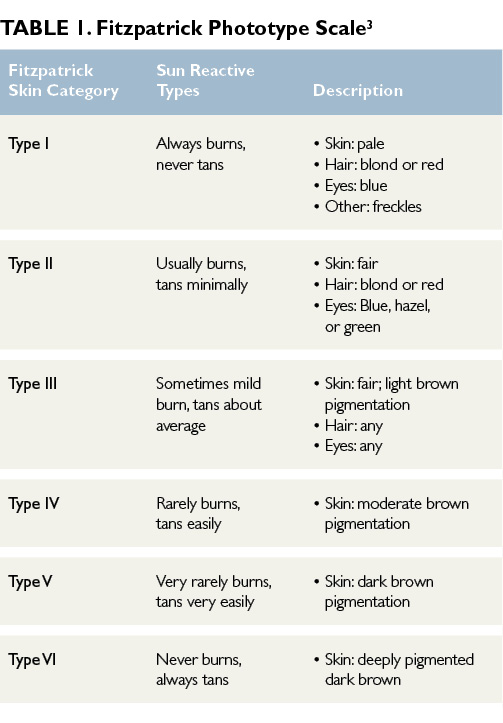
Actinic keratoses (AKs) are one of the most frequently encountered skin lesions in clinical practice.1,2 AKs are precancerous, focal, sun-induced areas of abnormal proliferation of atypical keratinocytes confined to the lower layer of the epidermis.1,2 Often referred to as solar keratoses, these lesions commonly occur in areas with chronic and cumulative sun exposure such as the face, ears, neck, dorsal forearms, hands, and scalp.1,2 The highest incidence of AK is seen in middle-aged to elderly men with chronic and sustained ultraviolet (UV) radiation exposure over their lifetimes. AKs are more prevalent in fair-skinned individuals and those categorized as Fitzpatrick skin types I and II (Table 1)3; the Fitzpatrick phototype scale is a numerical classification used to estimate the response of different types of skin to UV light.3

Click to enlarge

Etiology/ Histology

The main mechanism of AK formation is prolonged sun exposure and cumulative exposure to UV radiation.4 AKs result from the adverse effects of UV-A and UV-B light radiation on keratinocyte DNA.2,5 UV radiation induces cellular mutations and results in the formation of atypical keratinocytes.2,5 Mutations of genes may cause impaired apoptosis, uncontrolled cell proliferation, and progression to invasive squamous cell carcinoma (SCC).1,6 Although most AKs remain superficial and confined to the epidermis, some lesions will penetrate the reticular or papillary dermis and differentiate into SCC, especially when multiple AKs are present.1,6

Which of the following is the least effective topical therapy for actinic keratoses?

Clinical Presentation

The classic presentation of AKs is a gritty macule or small papule, varying in size, with an erythematous base and an overlying scaly white patch that is rough to palpation.2 Although AKs are commonly asymptomatic, patients may report occasional burning or lesion tenderness. Immunocompromised patients, specifically organ transplant recipients receiving treatment with immunosuppressive medications, are at increased risk of developing AKs.1,6 AKs may spontaneously regress, completely resolve, persist, or progress to SCC, particularly in the presence of multiple AKs.2 Clinically it is difficult to predict the course of AKs, and no method exists for predicting lesions that will evolve into invasive SCC.5 The risk of AKs transforming to SCC is minimal (1%)1; however, current guidelines suggest treating all AKs.4

Diagnosis

Diagnosis generally is made by visual inspection and palpation. Keratoacanthoma, SCC, basal cell carcinoma, and seborrheic keratosis lesions may have a similar presentation to AKs and must be considered in the differential diagnosis.2 Characteristic history and examination findings are usually adequate for diagnosing AK, but dermoscopy, if available, is recommended if the clinical exam findings are not typical of AK. A strawberry pattern, consistent with undulated vessels surrounding hair follicles filled with yellow keratotic plugs on an erythematous background, is characteristic of AK lesions on dermoscopy. Skin biopsy is performed if the clinical exam and dermoscopy findings are not typical of AK. Any clinically suspicious lesion — including lesions with increasing tenderness, rapid enlargement, or those recalcitrant to initial therapies — warrants skin biopsy for a definitive diagnosis.2     

Continue Reading Below

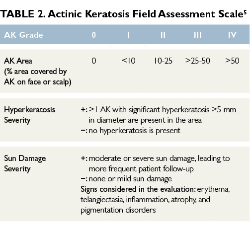
Surrounding the visible and palpable AKs lesions are skin areas prone to sun damage that are more likely to develop into clinical AKs or other sun-related cancers.5 Subclinical, nonvisible sun damage is known as field cancerization.5 Determining the severity of AKs and the extent of surrounding field damage can be challenging. Many AK grading tools use visible isolated lesion counts to assess the severity of the AKs, but they lack grading of the surrounding sun-damaged skin. The Actinic Keratosis Field Assessment Scale (AK-FAS) has been developed and tested as a relevant and reproducible assessment of AKs in clinical practice and aids in the grading of the severity of AKs and the field area damaged by UV radiation.5 Utilizing the scale can standardize a diagnosis, aid in selecting the most appropriate treatment, and provide an objective method to assess treatment response (Table 2).5

Click to enlarge

Read More

Identifying Best Candidates for Bexarotene Therapy in Cutaneous T-Cell Lymphoma: Role of APOA5, APOC3 Genotypes

Share this content:
The majority of the patients had mycosis fungoides, the most prevalent form of cutaneous T-cell lymphoma.

According to results of a retrospective, observational, noninterventionalist, multicenter Spanish case-series study designed to evaluate the role played by genetic polymorphisms in bexarotene-treated patients with cutaneous T-cell lymphoma (CTCL), screening of the APOA5 and APOC3 genotypes in these individuals may be a useful tactic for estimating triglyceride concentrations. Findings from the current analysis were published in JAMA Dermatology.

The investigators sought to examine the link between genetic polymorphisms of apolipoprotein genes APOA5, APOC3, and APOE, and to evaluate the severity of hypertriglyceridemia among patients with CTCL receiving bexarotene therapy, as well as to optimize patient selection for treatment with bexarotene, based on the expected adverse effect profile.

Continue Reading Below

The case series was performed at 12 university hospital dermatology departments in Spain between September 17, 2014, and February 17, 2015. A total of 125 patients with a confirmed diagnosis of CTCL who had received bexarotene for ≥3 months were enrolled in the study. Ultimately, 9 individuals were excluded because of missing analytic triglyceride data; thus, the final study group comprised 116 patients.

Data on demographic and cardiovascular risk factors were obtained for all participants. In addition, a complete blood analysis was performed, including a lipid profile, along with a genetic analysis from a saliva sample. The primary study outcome was the maximal triglyceride levels that were reported in association with the minor alleles of the polymorphisms being analyzed.

The mean patient age was 61.2±14.7 years; 59.5% of the participants were men, and 73.2% had mycosis fungoides, the most prevalent type of CTCL. While undergoing bexarotene treatment, 82.8% (96 of 116) of the participants developed hypertriglyceridemia, which was considered to be severe or extreme in 8.3% (8 of 96) of these patients.

Those individuals who carried minor alleles of the polymorphisms did not demonstrate any significant differences in baseline triglyceride levels. After bexarotene treatment, however, carriers of ≥1 of the 2 minor alleles of APOA5 c.1131T>C and/or APOC3 c.*40C>G had significantly lower concentrations of triglycerides compared with noncarriers of these minor alleles (241.59±169.91 mg/dL vs 330.97±169.03 mg/dL, respectively; P =.02), and were thus less likely to experience severe hypertriglyceridemia.

The researchers concluded that the findings from this study suggest that APO polymorphism investigation might prove beneficial in the clinical evaluation of bexarotene-induced hypertriglyceridemia in patients with CTCL and identification of the most suitable candidates for this treatment. Future studies aimed at exploring the sensitivity of these polymorphisms for predicting adverse outcomes to recognize which patients might need to discontinue treatment, or which will experience pancreatitis are warranted.

Follow @DermAdvisor

Reference

Cabello I, Alia P, Pintó X, e al. Association of APOA5 and APOC3 genetic polymorphisms with severity of hypertriglyceridemia in patients with cutaneous T-cell lymphoma treated with bexarotene. JAMA Dermatol. 2018;154(12):1424-1431.

Read More

Unique Presentation of SJS in a Female Patient Treated With TMP-SMX

This article originally appeared here.
Share this content:
Stevens Johnson syndrome (SJS) and toxic epidermal necrolysis (TEN) often presents 8–12 days after drug exposure

A recently published case report describing a 27-year-old female patient with a history of polycystic ovary syndrome discusses a rare presentation of Stevens Johnson syndrome (SJS) involving vaginal pain and mucocutaneous desquamation following re-exposure to trimethoprim-sulfamethoxazole (TMP-SMX).

The patient, who presented to the clinical office complaining of dysuria, was diagnosed with a urinary tract infection and was prescribed TMP-SMX. After taking 4 doses of her medication, she presented to the emergency department (ED) complaining of left labial swelling and pain. She was diagnosed with left vulvovaginitis and was told to discontinue TMP-SMX since her urine cultures were negative. Two days later, the patient returned to the ED complaining of worsening vaginal pain and vulvar lesions but was discharged with a sitz bath and supportive care and urged to follow-up at the clinical office. At her follow-up appointment, she was reinitiated on TMP-SMX due to  “concerns of vulvar cellulitis with possible underlying abscess.”

Continue Reading Below

“After she received 2 doses of TMP-SMX, the patient had new lip swelling, periorbital swelling, facial swelling, and formation of thick white plaque on her tongue and mouth,” the study authors reported. They added, “She also developed an erythematous papular pruritic rash on her hands, arms, soles of the feet bilaterally and papules were noted on the legs (up to thighs), feet, and abdomen.”

The study authors also noted that the patient experienced early vesicle formation, skin sloughing, significant erythema and edema in her genital region, a fever of 102°F, plaques on her tongue, and a painful maculopapular skin rash. The patient was diagnosed with SJS and transferred to the burn unit/ICU where she received intravenous immunoglobulin, supportive care, as well as wound care. The patient was discharged 11 days after being admitted to the hospital.

In their review, the study authors discussed several interesting features of this patient case, including the quick onset and development of SJS (6-12 hours vs 6 days to 2 weeks) as well as the unique presentation of the patient (vaginal pain/ mucocutaneous desquamation). They concluded, “This report shows that although SJS is a rare diagnosis, providers should consider SJS as a possible differential diagnosis as a cause of vaginal lesion after exposure to drugs.”

Follow @DermAdvisor

Reference

Mergler R, Chuang M; Stevens Johnson Syndrome with Vaginal Pain and Lesions as Initial Presentation. Am J Case Rep 2018; 19:1519-1521 DOI: 10.12659/AJCR.912123

Read More

Increased Risk for Incident Thyroid Disease in Psoriasis

Share this content:
An endocrinology consultation should be considered in patients with psoriatic disease who present with thyroid symptoms.

Patients with psoriatic disease have an increased risk for thyroid disease, including hyperthyroidism, hypothyroidism, thyroiditis, and certain autoimmune thyroid conditions such as Grave’s disease and Hashimoto’s thyroiditis, according to study results published in the Journal of the American Academy of Dermatology.

The investigators identified a total of 13,266 patients with psoriatic arthritis (the psoriatic arthritis group), 149,576 patients with psoriasis alone (the psoriasis group), and 162,842 controls who did not have psoriasis (the control group) from data collected between January 1, 2000 and December 31, 2012. Mean patient age in the psoriasis, psoriatic arthritis, and control arms was 45.11±20.09, 43.17±17.72, and 44.95±19.91, respectively (P <.0001); women comprised 46.21%, 37.83%, and 40.65% of the groups, respectively (P <.0001). A significantly higher percentage of the patients in the psoriasis and psoriatic arthritis arms had hypertension, type 2 diabetes, and hyperlipidemia compared with patients in the control group (P <.0001).

Continue Reading Below

Compared with the control group, the psoriatic arthritis and psoriasis groups had an elevated risk for incident hyperthyroidism (adjusted hazard ratio [aHR] 1.32; 95% CI, 1.07-1.65; P <.05 and aHR 1.22; 95% CI, 1.11-1.33; P <.0001, respectively) and Grave’s disease (aHR 1.38; 95% CI, 1.07-1.79; P <.05 and aHR 1.26; 95% CI, 1.131.41; P <.0001, respectively).

Moreover, both the psoriatic arthritis and psoriasis groups also had an increased risk for incident hypothyroidism (aHR 1.74; 95% CI, 1.342.27; P <.0001 and aHR 1.38; 95% CI, 1.231.56; P <.0001 respectively) and Hashimoto’s thyroiditis (aHR 2.09; 95% CI, 1.343.24; P <.01 and aHR 1.47; 95% CI, 1.181.82; P <.01, respectively) compared with the control arm.

A major study limitation was the lack of available data on psoriasis severity. In addition, in order to avoid overestimating the risk for incident thyroid diseases, patients with psoriasis diagnosed prior to 2000 were excluded from this study.

The investigators concluded that the findings demonstrate that an endocrinology consultation should be considered in patients with psoriatic disease who present with thyroid symptoms, particularly patients with psoriatic arthritis.

Follow @DermAdvisor

Reference

Wang S-H, Wang J, Lin Y-S, Tung T-H, Chi C-C. Increased risk of incident thyroid diseases in people with psoriatic disease: a cohort study [published online December 5, 2018]. J Am Acad Dermatol. doi:10.1016/j.jaad.2018.11.049

Read More